- What is cell division?
- Types of cell division
- Cell division in prokaryotes
- Eukaryotic cell division
- Chromosomes vs chromatids
- Cell division stages
- Mitotic cell division
- Meiotic cell division
What is Cell Division?
Cell division is the biological process by which a cell (mother cell) divides to produce at least two daughter cells. The mother cell can produce two identical daughter cells (mitosis or binary fission) or four daughter cells with different genetic material (meiosis).
Cell division is an essential process for the growth, health and reproduction of an organism. In multicellular organisms like humans, mitosis serves to restore the health of tissues by producing more cells to substitute old or damaged cells (although not all tissues can do this: neurons regenerate at a very limited rate and region of the brain). Meiosis, on the other hand, serves to create genetically variable gametes (sperm or egg cells) to maintain variability in the population when individuals reproduce via sexual reproduction.
Types of Cell Division
There are three types of cell division: mitosis, meiosis and binary fission. However, most of the time, when talking about cell division, we are referring to mitosis. Binary fission is exclusive of prokaryotes, whilst mitosis is characteristic of eukaryotes, and meiosis of eukaryotes that reproduce sexually (not asexually).
Cell Division in Prokaryotes
There is only one type of cell division in prokaryotes: binary fission.
Binary fission is the process by which bacteria reproduce asexually. Binary fission consists of the duplication of the bacterium's genetic material and the division of the mother cell into two daughter cells, each with a copy of the bacterium's DNA.
Even though mitosis and binary fission can seem similar, they are different processes:
- Binary fission is a form of asexual reproduction, whilst mitosis is a form of cell division used for the growth and repair of a single organism/tissue.
- Binary fission happens in prokaryotes and mitosis happens in eukaryotes.
- In binary fission, there is no mitotic spindle and the duplication of the genetic material happens at the same time as the separation of said genetic material into the two daughter cells. In mitosis, however, there is a separate phase (S-phase) for the duplication of genetic material and the chromosomes are separated into the daughter cells by the mitotic spindle.
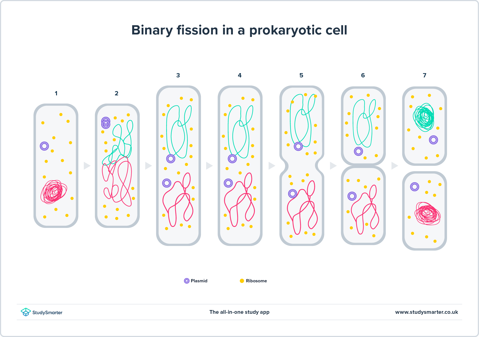
Fig. 1. Binary fission diagram. Note that there is no mitotic spindle in the image. Plasmids also count as genetic material in bacteria. They have been thought to distribute randomly in daughter cells, meaning that the copies of plasmids can vary between daughter cells. However, this random-distribution model has been called into question
1.
| Binary fission | Mitosis |
| Type of organism | Prokaryotes (bacteria) | Eukaryotes |
| Intent | Asexual reproduction | Growth and repair of an organism |
| S-phase (DNA replication phase) | Absent | Present |
| Mitotic spindle | Absent | Present |
The steps of binary fission are:
DNA replication (1-2 in figure 1)
Cytoplasm split: the cellular elements of the bacterium start to distribute into two compartments which will be the two daughter cells at the end of the division process (3-4)
Septum formation: a dividing wall forms between the two compartments, separating the cytoplasm and its content.
Cell constriction: the septum constricts, pinching the mother cell until it divides into two daughter cells (5-7).
Eukaryotic Cell Division
There are two types of cell division in eukaryotic cells - mitosis and meiosis.
- Mitosis produces two genetically identical daughter cells and is the most common type of cell division. Mitosis is fundamental to ensure the cells of the same organism carry the same genetic information (not accounting for mutations that happen spontaneously).

Fig. 2 -
An overview of mitosis. 2n - a diploid (two sets of chromosomes) cell- Meiosis produces four daughter cells that are genetically different, called reproductive cells or gametes like sperm or egg cells. Meiosis ensures variability in the offspring so that a species is not all clones of each other. Meiosis is a crucial process for sexually reproducing organisms.
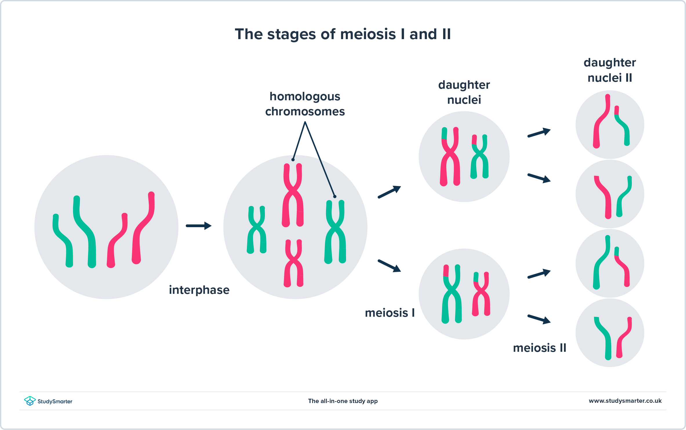
Fig. 3. The process of meiosis. As you can see, meiosis has two phases:
meiosis I and
meiosis II. The end product is four daughter cells, each with a -potentially- different genetic composition because they each receive a different combination of chromatids from the mother cell, which, additionally, might have exchanged genetic material before being pulled to the daughter cell.
Chromosomes vs Chromatids
To fully understand the differences between mitosis and meiosis, we need to understand the difference between chromosomes, chromatids and sister chromatids.
Chromosomes are x-shaped structures that contain DNA, i.e. the hereditary information passed on from the mother cell to the daughter cells during cell division. Chromosomes arise when the DNA in chromatin structure (which is less condensed and can be accessed for transcription) becomes more compact. They are present in the nucleus of eukaryotic organisms.
Prokaryotes do not have a nucleus, and it is also not correct to say they have chromosomes. They have a ring-shaped genophore (which contains DNA) instead.
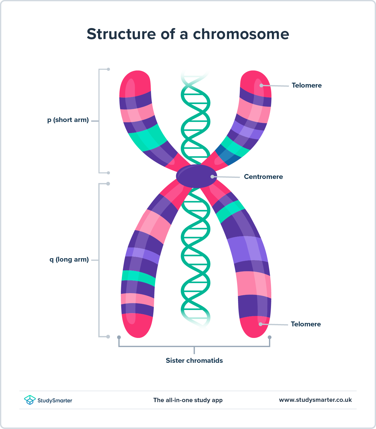
Fig. 4 - This is what a chromosome looks like. Telomeres are the ends of the chromosomes, and play a crucial role in keeping the chromosomes "healthy"
A chromatid is one of the DNA strands that makes a chromosome. One chromosome, therefore, has two chromatids, which are termed sister chromatids. Sister chromatids are held together at the centromere. The name "sister chromatid" comes from the fact that each strand of one chromosome (i.e. each of the two chromatids) contains exactly the same genetic information because they are exact copies of each other.
It's important to note that sister chromatids are those that are on the same chromosome, and should not be confused with chromatids that are on the homologous chromosome. Homologous chromosomes are those that carry the same genes but not the same alleles.
Humans are diploids, meaning that we have two copies of each gene, one from our mother and one from our father (i.e. one from the original egg cell and one from the original sperm cell that created us). These two copies, even though they carry information for the same genes (except for the sexual chromosomes X and Y, which have different genetic composition), they don't necessarily have the same sequence. In other words, they can code for different alleles of the same gene. The chromosomes that code for the same genes are called homologous chromosomes. This is a crucial concept in meiosis.
Therefore, each allele is carried in one chromosome. The unique combination of alleles each of us has is what makes all humans different.
When cell division is about to happen, the existing DNA duplicates, so now we have two copies of each allele: the sister chromatids. During cell division, the sister chromatids will be pulled away from each other to divide the genetic material between the daughter cells.
Cell Division Stages
Generally, when we ask about cell division stages, we are asking about the stages in mitosis and/or meiosis (not binary fission). These stages are more or less the same in both types of eukaryotic cell division, and share the same names, but they differ in some details. We will go over the basic names and functions of each stage here, and delve into the details in the following sections of the article.
Apart from cell division, cells have a whole cell cycle, of which mitosis or meiosis are only a part. The stage before cell division is termed interphase and it also has phases within it. Cells don't have to be constantly dividing, and will actually spend most of their time in the interphase, although this varies by cell type.
The order of the cell division stages is:
- Prophase
- Metaphase
- Anaphase
- Telophase
- Cytokinesis
Cell Division Stages - Prophase
During prophase, the nuclear envelope dissolves and chromosomes start to condense and become visible. Centrioles (microtubules found near the nuclear membrane) start to move to the opposite poles of the cell. The spindle fibres also start to form, which will later separate the chromosomes (meiosis I) or sister chromatids (mitosis and meiosis II).
Spindle fibres are a specialised cytoskeleton structure that forms during cell division to pull the chromosomes or chromatids to opposing sides of the cell.
Importantly, during metaphase is when the homologous recombination, characteristic of meiosis I, happens.
Cell Division Stages - Metaphase
In this stage, the chromosomes align along the middle of the cell, known as the metaphase plate, and the spindle fibres attach to the chromosomes at the kinetochores/centrosomes.
Kinetochores are protein structures that form where sister chromatids unite (sister chromatids are perfect copies of each other) and where the spindle fibres attach during cell division to separate the sister chromatids or homologous chromosomes. Kinetochores attach to the chromosomes in a specific region called the centromere.
Cell Division Stages - Anaphase
In the anaphase, sister chromatids (meiosis II and mitosis) or homologous chromosomes (meiosis I) are pulled to opposite ends of the mother cell by the spindle fibres.
Cell Division Stages - Telophase
During this stage, the chromatids or chromosomes reach opposite poles of the cell. The DNA starts to unwrap (from the chromosome structure it goes back to the chromatin structure) and a new nuclear envelope forms to surround it and reconstruct the nucleus of the cells.
Cell Division Stages - Cytokinesis
This is the final stage of cell division, where the two daughter cells are actually formed: the cytoplasm and cell membrane divide into two to generate the new cells. In plant cells, apart from the cytoplasm and the cell membrane, the cell will have to generate new cell wall to split the daughter cells. If the cell division process is meiosis, a new cell division round will have to happen: meiosis II.
Although cytokinesis is considered to be part of cell division, it is not strictly a part of mitosis or meiosis. Also, although it is explained as a different part of cell division, it usually happens at the same time as telophase.
Mitotic Cell Division
Mitosis is the division of somatic (body) cells. Mitosis does not take place in the production of sex cells (gametes). Mitosis, although a continuous process, to simplify, can be divided into the four main stages we mentioned above (prophase, metaphase, anaphase and telophase), plus cytokinesis at the end.

Fig. 5 - An overview of mitosis. 2n - a diploid (two sets of chromosomes) cell
The crucial thing to remember about the mitotic division process is that the end product is two genetically identical daughter cells, and cell division only happens once. For that to be the case, during the metaphase, all chromosomes, homologous and non-homologous, are set in line across the metaphase plate. During the anaphase, the sister chromatids are pulled to opposite sides of the cell.
Meiotic Cell Division
Meiosis and mitosis are different processes. However, the overall phases are the same: prophase, metaphase, anaphase, telophase and, at the end, also cytokinesis. Importantly, in meiosis the sequence of these phases happens two times: meiosis I and meiosis II.
Meiosis happens in sex cells. The development of sperm is called spermatogenesis, and the development of an egg is oogenesis. Spermatogenesis in males will continue throughout their lives, while oogenesis in females will only occur until menopause.
Most of the other animals will not go through menopause like human females. Only killer whales and pilot whales have been observed to do so. Other animals’ reproductive organs will last about as long as they will.
| Mitosis | Meiosis |
| Intent | Growth and tissue repair | Sexual reproduction (gamete production) |
| Types of mother cells | Somatic cells | Sex cells, gamete precursors |
| Number of daughter cells | Two, genetically identical | Four, genetically variable |
| Ploidy of daughter cells | Diploid, like the mother cell (2n) | Haploid, half the genetic material as the mother cell (n) |
| Genetic variability | No variability | Genetic variability is the goal. It is achieved by the process of recombination and random segregation of sister chromatids |
| Stages of cell division | Just one cell division | Two cell divisions: meiosis I and meiosis II |
Meiosis I
The first time the cell divides for meiosis, the daughter cells will have two sets of chromosomes (diploid), same as in mitosis. Unique to meiosis I, paired homologous chromosomes (bivalents) will go through homologous recombination (also known as crossing over) in which they exchange genetic information with each other. Homologous chromosomes (same genetic information) will line up and swap segments during prophase I.
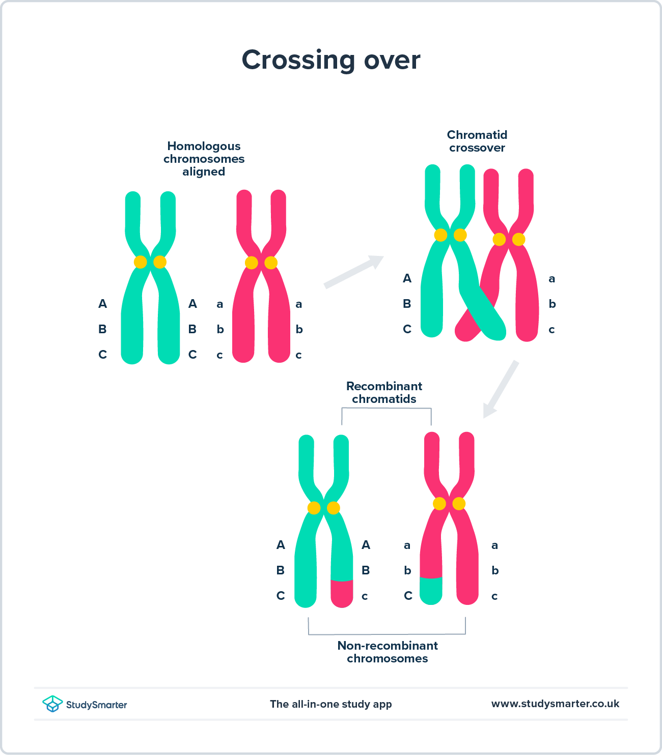
Fig. 6. Simple diagram of homologous recombination. Note that even though the segments of the chromosomes that are being exchanged code for the same genes, they do not have the exact same DNA sequence and code for different alleles, as represented by capital vs lowercase letters.Then, during anaphase I, instead of the sister chromatids being pulled to opposing sides of the cells, it will be whole homologous chromosomes that are separated. In other words, for each chromosome, one daughter cell will have two copies of the same alleles (two sister chromatids) and the other daughter cell will have the other two copies (the other two sister chromatids). Therefore, after cytokinesis, meiosis I produces two haploid daughter cells, with only one chromosome of each.
However, because of the homologous recombination of prophase I, the sister chromatids might not actually be 100% the same: genetic information (segments of DNA) were exchanged during that process, which increases variability in the offspring, as alleles are swapped and the chromosomes are no longer exactly like the ones inherited by the person from their mother and father.
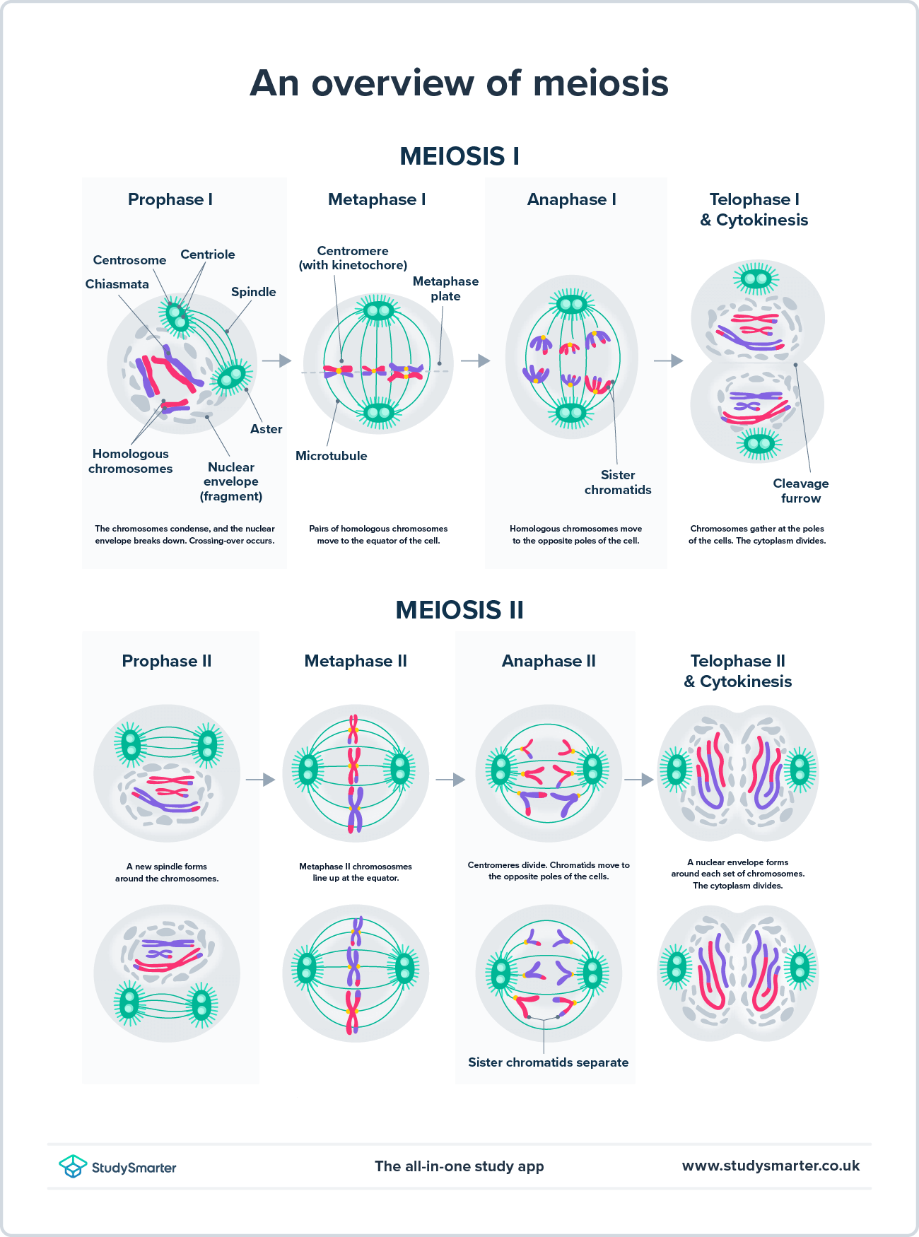
Fig. 7 -
An overview of meiosis. 2n - a diploid cell, n - a haploid (one set of chromosomes) cellFigure 3. An overview of mitosis. 2n - a diploid (two sets of chromosomes) cellMeiosis II
The second time the cell divides, the sister chromatids of the two daughter cells will split, and each will form two haploid (single-set of chromosomes) cells (also known as granddaughter cells).
There is an importance to egg and sperm cells being haploid. Male and female gametes will meet, and sperm will fertilise the egg. The embryo will contain two sets of chromosomes during fertilisation, becoming diploid. If the gametes were already diploid, the embryo would be tetraploid, so it's important that the DNA load is reduced by half during meiosis to avoid chromosomal mutations in the embryo.
The principle of meiosis II is almost identical to that of meiosis I. The two differences are:
Meiosis I will produce two haploid cells, and meiosis II will produce four haploid cells.
Genetic recombination only occurs in meiosis I.
During anaphase II, "sister" chromatids (remember that they might not be identical due to the recombination in prophase I) are pulled to opposite sides of the cell, like in mitosis. In meiosis I, it was homologous chromosomes that were pulled to each pole of the cell.
 Fig. 8 - An overview of meiosis II. Note how from a diploid mother cell we get two haploid daughter cells
Below you can find a table with the basic differences between mitosis and meiosis. To delve deeper into these cell division processes, you can visit our articles on the topic: Mitosis, Meiosis, Meiosis I, and Meiosis II.
| Meiosis I | Meiosis II |
| Mother cell | Diploid (2n) | Haploid (n) |
| Daughter cells | Haploid (n) | Haploid (n) |
| Homologous recombination | Yes, in prophase I | No |
| Anaphase I | Homologous chromosomes are pulled to opposing sides of the cell | Sister chromatids are pulled to opposing sides of the cell |
Importantly, during meiosis, from a
single diploid (2n) mother cell, we end up with
four haploid (n) daughter cells.
Cell Division - Key takeaways
Cell division of eukaryotic cells is either mitotic (somatic cells) or meiotic (sex cells). Unicellular cell divisions are for reproductive purposes such as budding in yeast and binary fission in bacteria.
Mitosis has four main stages - prophase, metaphase, anaphase and telophase. The cell will then physically split by cytokinesis. Mitosis produces two genetically identical cells.
Meiosis has two parts - Meiosis I and Meiosis II.
Both meiosis parts have the same stages as in mitosis. However, meiosis will produce four genetically different haploid cells.
References
- https://www.ncbi.nlm.nih.gov/pmc/articles/PMC4163524/
Similar topics in Biology


