Physics of vision: light and image capture
Light is composed of photons, which can behave as particles or waves. Each photon has a frequency that corresponds to the energy of the photon. The process by which photons form an image in our brain is quite complex, but we can break it down as follows:
- Photons are emitted by the Sun. They travel through space and enter the atmosphere where they impact objects as they pass through. Photons with higher frequencies have higher energies, and photons with shorter frequencies have smaller energies.
- When photons impact an object, they are reflected. Depending on the structure of the atoms in the object, some photons will be reflected and others will be absorbed. The colours we see are the photons reflected by the objects.
- The photons reflected with certain energies enter our eyes through the cornea, which the lenses inside our eyes will focus.
- The lenses project the ray of light made of photons onto the back of the eye.
- Finally, the photons will excite special cells known as cone cells or rod cells, each one responding to a different range of photon energies. These ranges are interpreted as colours in our brain.
Physics of the eye and vision
Below we look at the structure of the human eye and how we can see.
The eye
The eye is a complex organ composed of lenses, muscles, nerves, and tissue. It captures incident light and converts it into an electrical signal that is sent to the brain. Here are some of the main parts of the eye:
- Iris. This is the area around the eye’s aperture, and it has a colour depending on genetic factors. The area closes or opens to allow more or less light to enter the eye.
- Lenses. The lenses are a semitransparent structure located directly behind the main aperture of the eyes. Their unique function is to concentrate the rays of light to modify the focus of images at different distances. Refraction is also present in lenses.
- Cornea. This is the front, crystalline part of the eye that allows light to enter.
- Pupil. The black part located in the centre of the eye is the pupil. It is the aperture from which light enters.
- Vitreous humour. This gel-like substance fills the eye and is found between the lenses and the retina.
- Ciliary body and muscle. The ciliary muscle is responsible for changing the size of the focus of the eye when we focus on a near object. The ciliary body, which contains the ciliary muscle, produces a liquid named aqueous humour. The aqueous humour retains the internal pressure of the eye and transports nutrients.
- Retina. The retina is the surface behind the eye that is responsible for sensing, and it is filled with cone and rod cells.
- Optical nerve. This is the nerve that sends the signal to the brain.
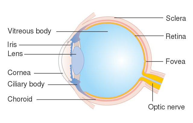
The structure of the human eye, Cancer Research UK CC BY-SA 4.0
Physics of Vision
Let’s briefly study how we are able to see!
Controlling the amount of light
A certain amount of light is needed in order for us to see. An aperture called the pupil regulates the amount of light entering the eye. This aperture is circular, and you can see it as black when you look at your eyes in the mirror.
The radius of the aperture is regulated by the iris, which is connected to the ciliary muscle. The muscle will expand and contract depending on the light conditions. When the intensity is higher, the muscles will contract, and when it is lower, the muscles will expand.
Forming a focused image
Light entering the eye passes through the cornea. The cornea will alter its shape depending on the object’s distance. In this case, when the ciliary muscle contracts, it changes the shape of the lenses, and this will change the distance at which we can focus an object.
Sensitivity of the eye
The photodetector in the eye is the retina. There are around 130 million light-sensitive cells in it. Each cell reacts to light from a single small point. A photopigment is a substance found in light-sensitive cells. The photopigment molecules absorb light and excite the cell, resulting in an electrical impulse that travels through a network of neurons to the optic nerve and, ultimately, the brain.
There are two main types of light-sensitive cells: rods and cones.
- Rods are specialised cells that help us see in the dark. They are sensitive to a wide range of wavelengths in the spectrum. They also have a low resolution and are much more sensitive to the incoming photons than cone cells. Despite this, they produce white and black colours.
- Cones are specialised cells that come in three types: red, green, and blue cone cells. Each cell type responds to a different range of light frequencies (wavelengths). They can also be divided into L-cones (sensitive to red and yellow), M-cones (sensitive to green), and S-cones (sensitive to blue).

A conceptual illustration of the basic structure of rod cells and cone cells in the eye, Wikimedia Commons
The human eye can capture images in both dark and bright environments. The pupil opens to around 8mm in low light and contracts to 1.5mm when exposed to higher luminosity.
Colour vision
Rod and cone cells can detect electromagnetic waves with wavelengths ranging from 380 nanometres to 750 nanometres (nm). Rod cells are sensitive to nearly the whole wavelength range, with a peak response at 500nm. Cone cells, on the other hand, contain one of three photopigments, each with a particular frequency/wavelength range of response with its own peak and reception.
One of these photopigments in a cone cell may absorb a photon of light when it reaches the retina. The wavelength of the light influences the likelihood of absorption. A certain wavelength will excite one of the three types of cone cells in a specific ratio. This allows the brain to recognise wavelengths and generate the colour sense.
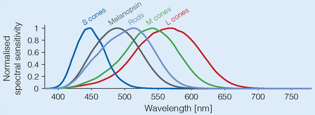
Overview of retina photoreceptors in the human eye, C. Blume, C. Garbazza & M. Spitschan, CC BY 4.0
Spatial resolution
The spatial resolution of an eye is its ability to see an object in greater detail. In the human eye, this resolution equals about one arc per minute, which is one degree between 160. This resolution allows us to see objects at one kilometre, which are separated by no less than 30cm. If we use the analogy of a camera, the overall resolution of the human eye is more than 576 megapixels per eye.
Physics of Vision - Key takeaways
- Vision in the eye is possible due to the photons emitted by the Sun. After the photons are emitted, they impact diverse objects, and in their interactions, part of the photons are reflected and partly absorbed. The frequency of the photons reflected is what makes the colours we see.
- The eye is a complex organ. Its parts include the pupil, iris, lenses, cornea, retina, optical nerve, ciliary muscle, ciliary body, and vitreous humour.
- There are two main types of light-sensitive cells: rods and cones.
- Rods are specialised cells that help us see in the dark. They produce white and black colours.
- Colour vision is possible thanks to the different sensitivity of cone cells. Cone cells are divided into three types with a different sensitivity range, and the brain uses each range to interpret colours.
Images1. Diagram showing the parts of the eye. https://commons.wikimedia.org/wiki/File:Diagram_showing_the_parts_of_the_eye_CRUK_326.svg
2. Overview of the retina photoreceptors. Spectral sensitivities of the photoreceptors in the human eye. https://commons.wikimedia.org/wiki/File:Overview_of_the_retina_photoreceptors_(b).png


